
 Check out our appearance on the BBC, showcasing the use of our medical device at Worcestershire Royal Hospital
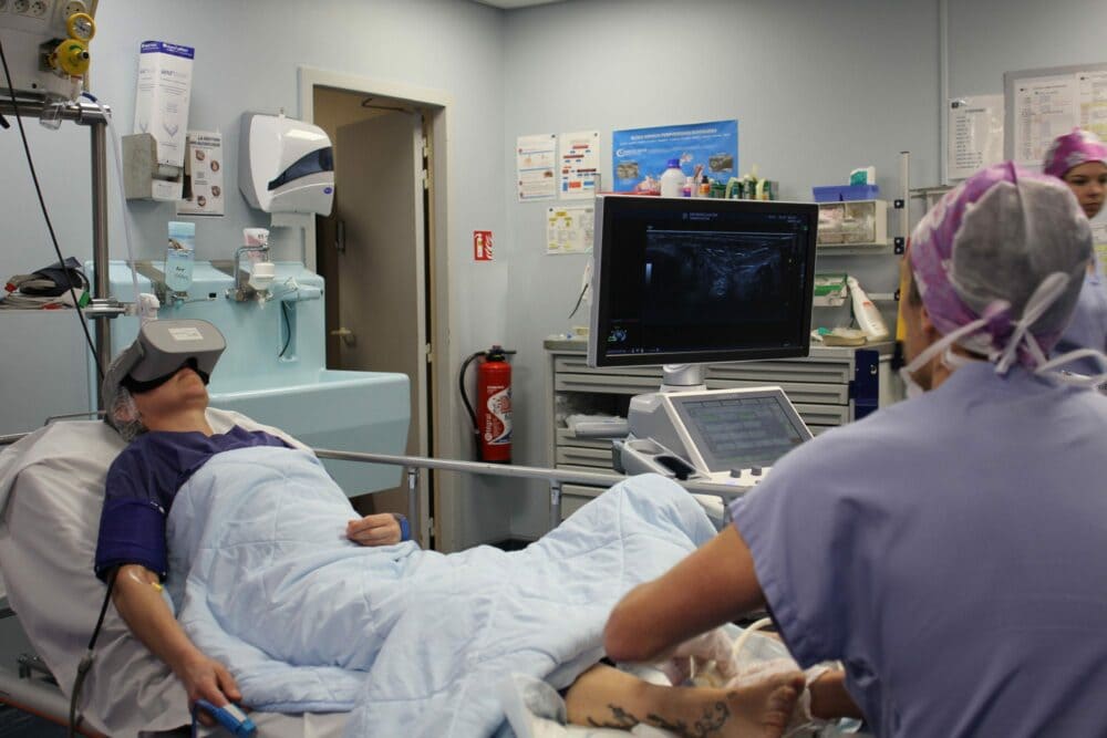
Relaxing during a medical imaging examination: it’s possible with virtual reality!
In France, 73.6 million imaging procedures were carried out in 2019 according to the French Institute for Patient Experience. CT scans, X-rays and ultrasounds are all essential examinations for adapting treatment and medical care. Although they are routine hospital procedures, this is not always the case for patients, who are confronted with the worry of the diagnosis and other anxiety-provoking elements. So how do you relax before or during a medical scan? In this article, we will first look at the specifics of the different examinations, in order to better understand where patients’ stress comes from. Finally, we will discuss the advantages of virtual reality to relax during medical imaging.
What are the different types of medical imaging?
Medical imaging can be divided into different types depending on the technique used: X-rays, ultrasound, magnetic field, natural or artificial radioactivity.
X-rays
The first medical imaging technique that we will discuss is frequently seen in orthopaedics, rheumatology and orthodontics. X-rays are a helpful examination to study bone trauma, skeletal deformities and dental implants. This procedure is also used to observe abnormalities in certain organs, such as lung or breast tumours (in the last case, mammography is the term used).
Depending on their density, tissues absorb the X-rays emitted to a greater or lesser extent and print a photographic film placed opposite the patient. This process is based on the same principle as photo film. Hollow structures such as the digestive system or joints can become opaque as a result of the injection of a contrast medium. According to this principle, the visualisation of blood vessels is called angiography and the observation of coronary arteries is called coronary angiography.
In order to reduce the dose of radiation, electronic detectors have replaced photographic film and are now used to digitise the images directly. X-rays are not a painful examination, but the pathology monitored by the images may cause suffering.
The CT scan
While radiography photographs dense structures in 2D, the scanner allows for 3D observation. This is the result of advances in computer technology and digital image processing, which led to the development of the computerised tomography (CT) scan in 1972.
Like X-rays, CT scans use X-rays to observe the human body. However, the resulting three-dimensional images look more realistic and are in the form of slices. A contrast medium may also be administered to study certain organs.
During a CT scan, an X-ray tube rotates around the patient at very high speed. During this rotation, images of the body are taken at 360°. This medical imaging makes it possible to identify a change in volume or a structural abnormality, whether it be an infection, a haemorrhage, ganglions or an embolism. In oncology, for example, the scanner checks the patient’s response to chemotherapy.
It is also used to guide drainage and biopsies. Medical imaging itself does not cause any painful sensations, but a procedure of this type may cause discomfort.
Ultrasound
Ultrasound is no longer based on the emission of X-rays, but on the use of sound waves that are imperceptible to the human ear: ultrasound. When ultrasound is emitted in the direction of a solid object, it bounces off it before returning to the starting point. Based on this principle, ultrasound measures the time required for this journey and produces a real-time image that shows the various structures.
This medical imaging technique is used to explore the heart, digestive, urinary and genital organs. Pregnancy ultrasounds are also based on this principle and reveal the environment, morphology and vitality of the foetus. Ultrasound has the advantage of being non-irradiating, painless and does not require the injection of a contrast medium.
Magnetic resonance imaging (MRI)
If there is any doubt after an X-ray, CT scan or ultrasound, the medical team turns to magnetic resonance imaging (MRI). During this examination, a magnetic coil is placed around the patient and scans the area of the body being studied, creating a magnetic field. MRI will allow the visualisation of different structures, including ‘soft’ tissues such as the brain, spinal cord, muscles, viscera or tendons.
MRI is an anxiety-provoking examination for some patients suffering from claustrophobia, as the inside of the machine can give a feeling of confinement. In addition, metal devices must be removed to perform this medical imaging.
Positron emission tomography (PET) and scintigraphy
Scintigraphy and positron emission tomography (PET) were developed after the discovery of radioactivity which led to the development of nuclear medicine. This invention of the 1990s consists of administering a molecule coupled to a radioactive element intravenously and following its evolution in the body. This tracer is non-toxic for the patient.
This type of medical imaging is used to explore the thyroid, the skeleton, cardiology and cancer. It thus provides information on function, but does not reveal anatomical data. Although the description of this examination may be impressive, it’s a painless procedure.
What are the stress factors during medical imaging?
As we have just seen, except in the case of interventional radiology, medical imaging examinations do not produce painful sensations. However, they still generate significant anxiety for some patients, to the point of disrupting the course of the examination or medical treatment.
One study found that most patients surveyed, about 91%, had a high level of anxiety while waiting for an X-ray examination. This research reveals that the stressful state experienced can have a physiological impact on the results, and thus lead to complications or misdiagnoses. Furthermore, a person who feels panic may decide to leave in the middle of an examination or not to carry it out, which has a major influence on the course of care and medical management.
During a PET/CT scan, the medical team injects radiopharmaceuticals, which results in a waiting period before the scan is performed. Although patients are told to relax, the reality is often different, as shown by the study conducted by researchers from the Nuclear Medicine Department of the Antoni Van Leeuwenhoek Cancer Institute (NKI-AVL) and scientists from Phillips Research Laboratories: mental and physical anxiety increases with the wait.
The effectiveness of the process can also be compromised if the patient is highly anxious. SuAzio Consulting’s Patient Impact on MRI Efficiency survey shows that anxious patients move around more, resulting in reduced image quality in 74% of cases.
Reducing patient stress prior to medical imaging is therefore necessary for both patient comfort and diagnostic efficiency. How can this be achieved?
The benefits of therapeutic virtual reality to relax during medical imaging
1. A multi-sensory experience for a sense of escape
Waiting periods, before or after medical imaging, present their share of stress for patients who are apprehensive about a still uncertain diagnosis. Anxiety mounts as one scenario follows another. In addition, the hospital environment isn’t always conducive to a sense of relaxation. To get out of this anxiety-provoking world, virtual reality offers therapeutic walks that have only one goal: to reduce this feeling of oppression and nervousness.
To achieve this feat, the Healthy Mind device uses hypnotic speech, a soothing landscape, music therapy and cardiac coherence to immerse the user in a safe and comfortable universe. Before and during examinations, the headset follows the patients. In the middle of a Zen garden or a sunny mountain, the wait seems more bearable and serene.
In order to maintain a pleasant feeling and to prevent the onset of headaches or nausea associated with virtual “motion sickness”, cyberkinetosis, the Healthy Mind software is designed on a teleportation principle. In this way, the destabilising factors of this technology are removed, leaving only deep relaxation.
2. Anxiety reduction without anxiolytics
We might be tempted to think that the use of a sedative would serve to reduce fear and anxiety before medical imaging. However, sedation is not always desirable as it involves more preparation and follow-up. On the other hand, some examinations require cooperation from the patient, for example to hold his or her breath or to maintain a particular position, all of which are incompatible with a loss of consciousness.
A study by Dr Anastos shows that introducing visual stimuli into an imaging environment reduces the need for sedation by up to 28%. Therapeutic virtual reality immerses patients in a relaxing landscape that isolates them from the anxiety-provoking elements of the examination. Moreover, a systemic study of 18 publications shows a significant reduction in stress and pain with a virtual reality headset. The use of such a device provides an anxiolytic effect without the use of sedative drugs and thus limits the need for anaesthesia.
3. Non-drug pain treatment
When we experience pain, we tend to focus on it. The sensation takes precedence over everything else and it becomes difficult to turn away from this focus. To help patients change their perspective, virtual reality headsets are proving very useful.
Therapeutic virtual reality is an effective device for diverting patients’ attention and transmitting a relaxing hypnotic speech. The immersion it offers is effective in the treatment of pain as it addresses the attentional resources of patients to escape from their painful experience and focus on another sensation.
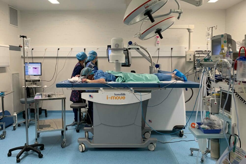
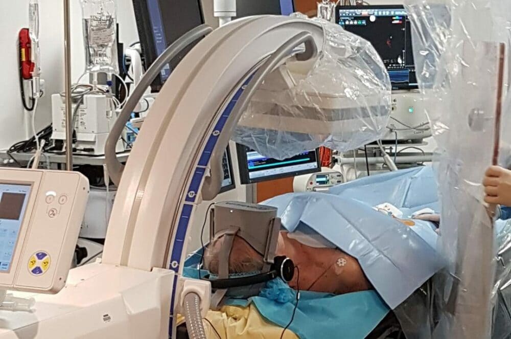
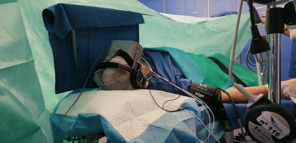
Although medical imaging examinations are generally painless, this is not always the case with interventional radiology. Therefore, VR headsets are used not only to ease patients’ anxiety, but also to reduce their painful sensations.
An attractive approach for many patients, as Chrystelle Hautecoeur, a nurse at the Eure-Seine hospital centre, explains:
“The virtual reality glasses have enabled a modification of the painful perception for several patients. (…) Its use has given confidence to patients looking for non-medicinal means of pain management.”
4. An ergonomic and non-invasive device
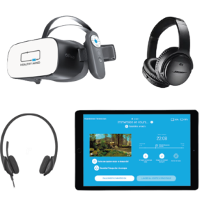
The virtual reality device consists of a wireless headset, headphones and a tablet. As a result, the tool is easy to carry and adjust. The absence of cables means that it can be adapted to changes in position and the constraints of the medical environment.
Furthermore, the Healthy Mind device offers personalised interactions throughout the immersion to maintain constant contact between the patient and the caregivers. The adjustable duration of the sessions makes the device practical as it adapts to all waiting times.
Due to the presence of metal elements, the virtual reality headset cannot accompany patients during an MRI. However, patients can still enjoy a moment out of time before going to the examination and feel more serene.
Therapeutic virtual reality offers a non-drug alternative to relax patients before, during and after medical imaging. This practical and ergonomic device adapts to all scenarios and provides a safe bubble in which to immerse oneself in case of anxiety. Due to its anxiolytic and analgesic effect, there are many use cases. If you would like to find out more about the device, we would be delighted to give you a demonstration.
Sources :
- Lo Re G, De Luca R, Muscarneri F, et al. Relationship between anxiety level and radiological investigation. Comparison among different diagnostic imaging exams in a prospective single-center study. Radiol Med. 2016 Oct;121(10):763-8. doi:10.1007/s11547-016-0664-z. Epub 2016 Jun 22.
- Grey SJ, Price G, Mathews A. Reduction of anxiety during MR imaging: a controlled trial. Magn Reson Imaging. 2000;18:351-55.
- Vogel WV, Valdés Olmos RA, Tijs TJ, Gillies MF, van Elswijk G, Vogt J. Intervention to lower anxiety of 18F-FDG PET/CT patients by use of audiovisual imagery during the uptake phase before imaging. J. Nucl. Med. Technol. 2012;40:92-98.
- Realizing productivity gains in MRI. Focusing more on the patient can boost the efficiency of MRI procedures. Survey commissioned by Philips and carried out by suAzio Consulting. 2017.
- Anastos, JP. The Ambient Experience in pediatric radiology. Journal of Radiology Nursing. 2007;26(2):50-55.